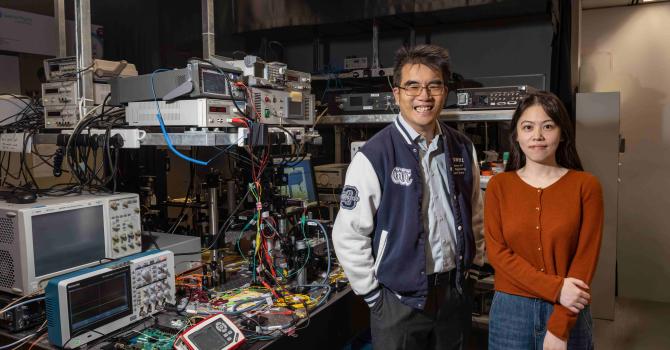
A research team led by Prof. Jianan QU and Prof. Kai LIU (LIFS) has developed long-term in vivo imaging technique to better understand and treat spinal cord injury
A research team led by Prof. Jianan QU and Prof. Kai LIU of Division of Life Science has developed an innovative technology for in vivo imaging of the important biological processes involved in the injury and repair of spinal cords, paving the way for a better understanding of the pathology and potential treatment of spinal cord injury (SCI).
“Considering the difficulties associated with long-term and repetitive spinal cord imaging, this innovation will be an important and widely used tool for the study of spinal cord injury,” said Prof. QU, who is an expert of optical engineering and science with extensive experience in in vivo linear and nonlinear optical spectroscopy and imaging of biological tissues from a variety of animal models.
The research paper was recently published in Nature Communications. The first author of the study is Wanjie WU, PhD student of Prof. Jianan QU.
The Paper
Nature Communications - Long-term in vivo imaging of mouse spinal cord through an optically cleared intervertebral window
Related Links
HKUST News – HKUST researchers develop long-term in vivo imaging technique to better understand and treat spinal cord injury
EurekAlert! – HKUST researchers develop long-term in vivo imaging technique to better understand and treat spinal cord injury
Study co-leads by Prof. Qu Jianan (right), Prof. Liu Kai (left), and the first author of the study WU Wanjie (second right), PhD student at Department of Electronic and Computer Engineering.
The multimodal molecular microscope system specially developed for the study.


